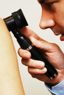
Dermoscopy
Dermoscopy has been shown to significantly improve the diagnosis of melanoma compared to standard clinical examination.
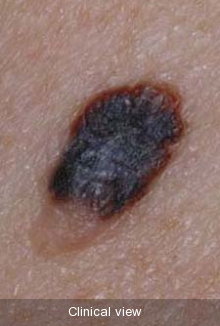
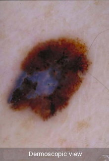
The hand-held dermatoscope eliminates surface reflection from the skin so that subsurface features can be seen. These features would otherwise not be visible - regardless of the magnification. Using dermoscopy, melanoma can demonstrate a number of features that are not seen in normal moles.
Dermoscopy is useful not just in the diagnosis of melanoma, but also in diagnosing many other types of skin spots and cancers, for example basal cell carcinoma.
It is important that a doctor performs dermoscopy at the time of consultation, and doesn’t just review recorded dermoscopic images at a later time. This is because there can be subtle features present that are lost when an image is recorded. For example, blood vessels may be compressed under the dermatoscope lens when an image is being recorded. If the melanoma is not producing pigment (amelanotic melanoma) then many of the usual clues to melanoma diagnosis will not be present, so assessment of the vessels then becomes crucial. Live dermoscopy will reveal these subtle features and is an important part of the diagnostic process.